Ultrasound imaging
Ultrasound is sound with frequencies above the range of human hearing, above about $20\ \mathrm{kHz}$. Medical ultrasound imaging typically uses frequencies between about $2\ \mathrm{MHz}$ and $15\ \mathrm{MHz}$.
A transducer held against the skin both sends and detects the ultrasound. Inside it, a crystal vibrates when a voltage is applied across it, and it produces a voltage when a returning wave makes it vibrate.
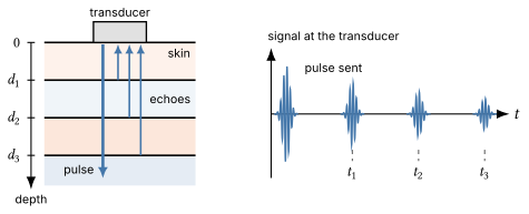
The transducer sends a short pulse of ultrasound into the body. Then it detects the echoes that return from inside the body. The machine uses the time each echo takes to return to find the depth of the boundary that reflected it.
Pulse-echo imaging
The pulse travels through soft tissue at about $1540\ \mathrm{m/s}$. At each boundary between two kinds of tissue, part of the pulse reflects as an echo, and the rest continues deeper. An echo from a deeper boundary takes longer to return.
Depth from echo timing
An echo from a boundary at depth $d$ travels down to the boundary and back up, a total distance $2d$. So it returns at time $t = 2d/v$ after the pulse is sent. The machine finds the depth of each boundary from the time its echo returns,
\[d = \frac{vt}{2}.\]It assumes that the speed of sound is $1540\ \mathrm{m/s}$ everywhere in the body.
The machine sends pulses along many closely spaced lines, one after another, to build up a two-dimensional image of the boundaries inside the body.
Reflection at a boundary
When sound reaches a boundary between two media, part of it reflects and the rest is transmitted. The property of each medium that matters is the product of its density and its speed of sound, called its acoustic impedance. The more the acoustic impedances of the two media differ, the larger the fraction that reflects.
| Boundary | Typical fraction of the intensity reflected |
|---|---|
| Fat and muscle | about 1% |
| Soft tissue and bone | about 40% |
| Soft tissue and air | about 99.9% |
Fractions are for sound that strikes the boundary straight on.
A boundary between two soft tissues reflects only a small fraction of the pulse. The rest continues deeper, so one pulse returns echoes from many depths.
A layer of air between the transducer and the skin would reflect almost all of the ultrasound. So the operator spreads a gel on the skin, which fills the gap and keeps air out.
Why megahertz frequencies?
An ordinary ultrasound image cannot show details much smaller than the wavelength of the ultrasound. The finest detail that an image can show is called its resolution, so shorter wavelengths give better resolution.
| Frequency | Wavelength in soft tissue |
|---|---|
| $20\ \mathrm{kHz}$ | $7.7\ \mathrm{cm}$ |
| $1\ \mathrm{MHz}$ | $1.5\ \mathrm{mm}$ |
| $5\ \mathrm{MHz}$ | $0.31\ \mathrm{mm}$ |
| $15\ \mathrm{MHz}$ | $0.10\ \mathrm{mm}$ |
The table uses $\lambda = v/f$ with $v = 1540\ \mathrm{m/s}$. Audible sound has wavelengths of centimeters or more in tissue, far too long to show the structure of an organ. Frequencies of several megahertz give wavelengths of a fraction of a millimeter.
Attenuation
As a pulse travels through tissue, the tissue absorbs part of its energy, which becomes thermal energy, and scatters part of it in other directions. So the intensity of the pulse decreases as it travels deeper. This decrease is called attenuation.
Attenuation increases with frequency. A higher-frequency pulse loses its intensity in a shorter distance, so its echoes from deep boundaries are too weak to detect.
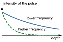
So the choice of frequency is a trade-off. Higher frequencies show finer detail but cannot image as deep. Structures just under the skin are imaged at higher frequencies, and deep organs at lower frequencies.
Example: Echo timing and wavelength
A transducer sends a pulse of $5.00\ \mathrm{MHz}$ ultrasound into the body. The echo from a boundary inside the body returns $52.0\ \mu\mathrm{s}$ after the pulse is sent. Use $v = 1540\ \mathrm{m/s}$ for the speed of sound in soft tissue.
- How far below the skin is the boundary?
- What is the wavelength of the ultrasound in the tissue?
Solution
1. The echo travels down to the boundary and back, so the depth is
\[d = \frac{vt}{2} = \frac{(1540\ \mathrm{m/s})(52.0\times 10^{-6}\ \mathrm{s})}{2} = 4.00\times 10^{-2}\ \mathrm{m} = 4.00\ \mathrm{cm}.\]2. The wavelength is
\[\lambda = \frac{v}{f} = \frac{1540\ \mathrm{m/s}}{5.00\times 10^{6}\ \mathrm{Hz}} = 3.08\times 10^{-4}\ \mathrm{m} = 0.308\ \mathrm{mm}.\]Doppler ultrasound
Measuring blood flow with ultrasound
Red blood cells scatter a small fraction of an ultrasound wave back toward the transducer. If the blood moves toward or away from the transducer, the echo’s frequency is Doppler-shifted, and the machine finds the speed of the blood from the shift.
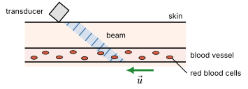
Only the part of the blood’s velocity along the beam shifts the frequency, so a beam perpendicular to the flow detects no shift. The operator tilts the transducer and marks the direction of the flow on the image, so the machine can correct for the angle. In our calculations, the blood moves directly toward or away from the transducer.
The echo is shifted twice
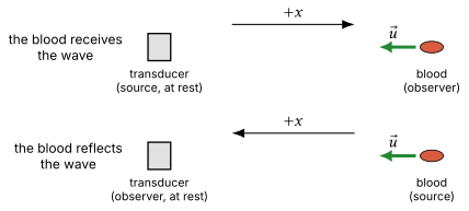
Let the transducer, at rest, send ultrasound with frequency $f_0$, and let the blood move directly toward the transducer at speed $u$. The Doppler shift happens twice, so we use the Doppler equation twice.
The frequency of the echo
First, the blood is a moving observer. With the $+x$ direction pointing from the transducer toward the blood, $v_{S,x} = 0$ and $v_{O,x} = -u$, so the blood receives the frequency
\[f_1 = \frac{v + u}{v}\,f_0.\]Then the blood reflects the wave, so it acts as a moving source with frequency $f_1$. Now the $+x$ direction points from the blood toward the transducer, so $v_{S,x} = +u$ and $v_{O,x} = 0$. The transducer detects the frequency
\[\begin{aligned} f_{\text{echo}} &= \frac{v}{v - u}\,f_1\\ &= \frac{v + u}{v - u}\,f_0. \end{aligned}\]Both shifts increase the frequency, because the blood moves toward the transducer in both steps.
The size of the shift
The frequency shift of the echo is
\[\begin{aligned} \Delta f &= f_{\text{echo}} - f_0\\ &= \Big(\frac{v + u}{v - u} - 1\Big) f_0\\ &= \frac{2u}{v - u}\,f_0. \end{aligned}\]Blood moves far more slowly than sound travels in tissue, $u \ll v$, so we can replace $v - u$ with $v$. This gives
\[\Delta f \approx \frac{2u}{v}\,f_0.\]The shift is proportional to the speed of the blood.
If the blood moves away from the transducer, the echo returns with a lower frequency. So the shift gives the direction of the flow as well as its speed.
Hearing the blood flow
For a transducer frequency of $5.00\ \mathrm{MHz}$ and blood moving toward the transducer at $0.500\ \mathrm{m/s}$, the frequency shift is
\[\Delta f \approx \frac{2(0.500\ \mathrm{m/s})}{1540\ \mathrm{m/s}}\,(5.00\times 10^{6}\ \mathrm{Hz}) = 3.25\ \mathrm{kHz}.\]The frequency of the echo differs from $f_0$ by less than 0.1%. But the shift itself, about $3\ \mathrm{kHz}$, is a frequency in the range of human hearing. Doppler ultrasound machines can play the shift through a speaker, so the operator can hear the blood flow.
Example: The speed of blood in an artery
A Doppler ultrasound transducer sends ultrasound with a frequency of $4.00\ \mathrm{MHz}$ toward an artery. The echo from the blood returns with a frequency $1.30\ \mathrm{kHz}$ lower than the frequency the transducer sends. Assume that the blood moves directly toward or away from the transducer, and use $v = 1540\ \mathrm{m/s}$ for the speed of sound in soft tissue.
- Is the blood moving toward or away from the transducer?
- How fast is the blood moving?
Solution
1. The echo’s frequency is lower than $f_0$, so the blood moves away from the transducer.
2. For blood moving away, the signs flip in both steps, and
\[f_{\text{echo}} = \frac{v - u}{v + u}\,f_0.\]The shift has magnitude $2u f_0/(v + u)$, which is about $2u f_0/v$ because $u \ll v$. Solving for the speed,
\[u = \frac{v\,\lvert\Delta f\rvert}{2f_0} = \frac{(1540\ \mathrm{m/s})(1300\ \mathrm{Hz})}{2(4.00\times 10^{6}\ \mathrm{Hz})} = 0.250\ \mathrm{m/s}.\]The exact form, $u = v\lvert \Delta f \rvert/(2f_0 - \lvert \Delta f \rvert)$, also gives $0.250\ \mathrm{m/s}$.
Example: Echolocation
A bat navigates by echolocation. It emits ultrasound and listens for the echoes. The bat flies directly toward a wall at $15.0\ \mathrm{m/s}$ while emitting ultrasound at $40.0\ \mathrm{kHz}$. What frequency does the bat detect in the echo from the wall? Use $v = 343\ \mathrm{m/s}$.
Solution
First, the bat is a moving source, emitting $f_0 = 40.0\ \mathrm{kHz}$, and the wall is an observer at rest. The wall receives the frequency
\[f_{\text{wall}} = \frac{v}{v - 15.0\ \mathrm{m/s}}\,f_0 = \frac{343}{328}\,(40.0\ \mathrm{kHz}) = 41.83\ \mathrm{kHz}.\]The wall reflects $f_{\text{wall}}$ as a source at rest, and the bat is an observer moving toward it. The bat detects
\[f = \frac{v + 15.0\ \mathrm{m/s}}{v}\,f_{\text{wall}} = \frac{358}{343}\,(41.83\ \mathrm{kHz}) = 43.7\ \mathrm{kHz}.\]In one step,
\[f = \frac{v + 15.0\ \mathrm{m/s}}{v - 15.0\ \mathrm{m/s}}\,f_0 = \frac{358}{328}\,(40.0\ \mathrm{kHz}) = 43.7\ \mathrm{kHz}.\]The small-speed approximation $\Delta f \approx (2u/v) f_0$ gives $43.5\ \mathrm{kHz}$. Here $u/v = 0.044$ is too large for the approximation to hold to three significant figures.
Summary
An ultrasound machine finds the depth of each boundary inside the body from the time its echo takes to return, $t = 2d/v$. Shorter wavelengths show finer detail, so imaging uses megahertz frequencies. Higher frequencies are attenuated more strongly, so they cannot image as deep.
Doppler ultrasound measures the speed of blood. The echo from moving blood is shifted twice. For blood moving toward the transducer at speed $u$, $f_{\text{echo}} = f_0 (v + u)/(v - u)$, and the shift is $\Delta f \approx (2u/v) f_0$.